
 English

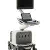


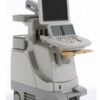
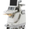
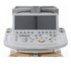
Philips iE33 – Refurbished Ultrasound
The iE33 xMATRIX system brings extraordinary clinical utility to every setting where echocardiography is used.
Περιγραφή
Philips iE33 – Refurbished Ultrasound
Not just a revolution, a revelation
This focus is what spurred the development of xMATRIX technology. Clinically advanced and thoughtfully designed, the iE33 xMATRIX system brings extraordinary clinical utility to every setting where echocardiography is used.While many technology companies call their innovations revolutionary, we know that innovation means nothing if it doesn?t help you. That?s why we won?t call the iE33 xMATRIX system a revolution, despite its cutting edge technology. Instead, we call it a revelation. It will change how you see ultrasound. More importantly, it will change what you see with ultrasound.
Clinical utility
The iE33 xMATRIX provides:? Flexible and intuitive workflow based around the user? Clear views of the cardiac anatomy, enabling accurate EF calculations? Live 3D volumes with excellent image quality and high frame rates – no more stitch artifacts with single beat? Fast and easy measurements of Live 3D and MPR views? More clinical information in one view with Live 3D and tools such as Dual Volume Display? A new approach to cardiac mechanics quantification using the next generation of SmartExam for echo studies? A simplified vascular exam with Auto Doppler, to assist with image and sample volume placementRanked #1 in 2012 Best in KLAS AwardsPhilips iE33 ultrasound system was ranked #1 among cardiovascular ultrasound systems in the 2012 Best in KLAS Awards: Medical Equipment & Infrastructure report. Described as an ?Independent Report from Healthcare Executives and Professionals,? the Best in KLAS Awards are based on evaluations from and interviews with healthcare executives, managers and clinicians from thousands of healthcare organizations, and reflect excellence in both service and product functionality. KLAS is an independent research firm specializing in monitoring and reporting performance of healthcare vendors.3D proves valueIn recent years, many studies have investigated the value of 3D imaging. Clinical experts have declared that it is both complementary and supplementary to 2D imaging. In particular, 3D echocardiography appears to improve accuracy and reproducibility over 2D echo in LV volume and function calculation, as well as the derivation of the mitral valve area in patients with mitral stenosis. 3D echocardiography is also becoming the standard of care in the interventional lab.
Innovations in image quality and workflow
You?ve come to expect image quality revelations from Philips. And with good reason. Philips innovations contribute to nearly every aspect of cardiac imaging. Now, we bring you a new revelation: changing imaging modes from 2D to 3D can be as easy as touching a button, without any compromise in image quality.3D without 3D hasslesAs the evidence mounts that volume imaging can provide highly relevant information, more and more clinicians are searching for a way to incorporate 3D imaging into their exams. The iE33 xMATRIX system removes the barriers to 3D imaging, giving clinicians the power to choose 2D, 3D or combination imaging without disrupting workflow. With the highly-ergonomic new X5-1 transducer, a simple push of a button brings Live 3D imaging to any exam.Focused on image qualityWhile much of the iE33 xMATRIX system is cutting-edge, we?ve retained all the imaging advantages that made the iE33 a best-in-class system:? PureWave Transducer Technology is the foundation for a host of imaging advances. The iE33 leverages PureWave crystal?s acoustic efficiency in transducer tuning and system optimization, facilitating imaging of a wide range of patient types with fewer artifacts and better penetration than conventional transducer technology. PureWave technology also enables miniature crystals, which require less stimulation to achieve high image quality, and make it possible to incorporate the power of xMATRIX in ergonomically sized transducers that can perform both 2D and 3D exams.? Advanced XRES performs 350 million calculations per frame of image data at rates of up to 500 frames per second, removing the noise from the LV cavity to make it easier to define the endocardial border.? Adaptive Broadband Flow uses the entire broadband frequency range within color Doppler, automatically adapting the frequency to the ROI to enhance spatial resolution. This is particularly useful when imaging the pulmonary vein, aortic insufficiency, and mitral regurgitation flow.Access data from your PACS on the iE33DICOM Query/Retrieve makes it possible to retrieve images from the PACS on the iE33 xMATRIX system. This feature enables easy comparison of past and current studies without use of a reading station. Users can choose to review previous studies before an exam, or use LiveCompare to view past studies in a loop while imaging the patient.
Επιπλέον πληροφορίες
| Βάρος | 150 κ. |
|---|---|
| Διαστάσεις | 56 × 110 × 140 cm |
| Brand | |
| Condition | |
| Medical Specialty | |
| Availability | |
| Color | |
| Doppler | CW Color Doppler, PW Color Doppler, Real-time Doppler Auto Trace |
| Display Technology | |
| Image Quality | |
| Screen Dimensions | |
| Touch Panel | |
| Connectivity | Analog Video Input/Output, DICOM, Ethernet, USB 2.0, DVD External, VGA |
| Print Type | Ethernet Color Printer, Thermal B/W Printer, USB Color Printer |
| Head ports | |
| System Portability |
Product Files
Product Files
Instruction Manual – SpecificationsDeclaration of Conformity
Compatible Probes
Compatible Probes
ConvexC5-1C5-2C8-5C9-4
LinearL9-3L11-3L17-5io
Phased ArrayS5-1S8-2
Independent CW DopplerD2TCCD2CWCD5CWC
TEES7-2omniS7-2
xMatrixX3-1X5-1X7-2TEE?X7-2t
