
 English

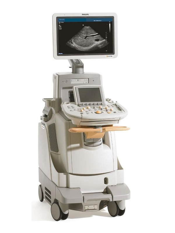


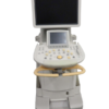
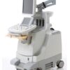
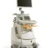
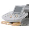
Philips iU22 – Refurbished Ultrasound
The iU22 intelligent ultrasound system continues to push the boundaries of image quality, ease of use, automated quantification, and throughput to help you address these challenges.
Περιγραφή
Philips iU22 – Refurbished Ultrasound
Keeping pace with growing requirements within the medical community and responding to patient interests are key challenges for today?s women?s healthcare providers. Diverse needs, department schedules, and tight budgets require efficiency and simplicity.The iU22 intelligent ultrasound system continues to push the boundaries of image quality, ease of use, automated quantification, and throughput to help you address these challenges.
Experience efficiencies with volume imaging
In a short time, volume imaging has become a readily accepted and expected part of many exams. And the good news is that it?s more than just a fetal face. Volume imaging provides you with new views and data that you could not acquire with conventional 2D imaging.Volume imaging is also more than just 3D and 4D. All the information you need is available in volume data sets. With the iU22 system, you can immediately and easily integrate volume imaging into your exams.How is it so simple? The iU22 system provides you with the ability to acquire volume data, interrogate it during the exam or later after your patient has been released, quantify it on-cart or off-cart, and easily transfer images and measurements to reports.Easy acquisitionThere are several methods of acquiring volume data. Freehand acquisition allows you to use virtually any transducer for advanced volumetric interrogation and display.The VL13-5, V6-2 and 3D9-3v automated volume mechanically steered arrays are easy to use and provide you with volume data that is quantifiable. Clinical uses include breast, gynecologic endometrial evaluation, fetal surface structural assessment, and fetal cardiac examination.The most advanced method of live volume acquisition is xMATRIX array. From one volume data set, you can generate an extraordinary number of images and anatomical views as well as perform advanced quantification.Remarkable visualization Because volume imaging has the potential to give you so much data, how are you sure you have the correct data? The iU22 system provides new tools to display and manipulate volume data sets.
Capture the moving target with STIC
Not all targets are still, especially in fetal cardiac imaging. The rapid movement of the fetal heart makes 3D imaging more challenging. That?s where Spatio-Temporal Image Correlation (STIC) adds value. STIC is a technology that uses the system?s ability to capture multiple volumes and synchronize them based on a calculated heart rate. The resulting volume image can be displayed and interrogated in real time. Using STIC, you can select various views for displaying the data to fully evaluate fetal heart anatomy and function.Find your best images with iSlice The QLAB iSlice feature was developed specifically for volume imaging. iSlice makes it easy for you to perform precision slicing of the volume to find the images with the best views and content for making your diagnoses. You can adjust the format to display 4, 9, 16 or 25 2D images based on slices from the volume set. You?ll quickly find that iSlice augments your diagnostic, decision making, and patient management processes.How will volumetric imaging impact your exams? A new use model shows the potential to improve your exam efficiencies by changing the way you acquire and visualize ultrasound data. When you integrate advanced volume methods, you can decrease the acquisition time of the exam because you are acquiring a few volume sets to complete the study, instead of 30 or 40 images. Volume imaging provides you with more data in less time.
Achieve new levels of image quality
You continue to tell us that achieving the absolute best image quality is critical to addressing your diagnostic challenges?every exam, every day. That?s why we?ve focused on transducer design and imaging technologies that increase the clarity and accuracy of data on all your patients.
- Revolutionary architecture
- Transducers designed for practical use
- Your solution to scanning technically difficult patients
- SonoCT image compounding and advanced XRES for superb tissue definition
- xMATRIX is the technology for the future
Επιπλέον πληροφορίες
| Βάρος | 150 κ. |
|---|---|
| Διαστάσεις | 56 × 110 × 140 cm |
| Brand | |
| Condition | |
| Medical Specialty | Cardiology, Neurosurgery, Obstetrics ? Gynecology, Radiology, Rheumatology, Urology |
| Availability | |
| Color | |
| Doppler | CW Color Doppler, PW Color Doppler, Real-time Doppler Auto Trace |
| Display Technology | |
| Image Quality | |
| Screen Dimensions | |
| Touch Panel | |
| Connectivity | Analog Video Input/Output, DICOM, Ethernet, USB 2.0, DVD External, VGA |
| Print Type | Ethernet Color Printer, Thermal B/W Printer, USB Color Printer |
| Head ports | |
| System Portability |
Product Files
Product Files
Instruction Manual – SpecificationsDeclaration of Conformity
Compatible Probes
Compatible Probes
ConvexC5-1C5-2C8-5C9-4
EndocavityC8-4vC9-5ecC10-3v
LinearL9-3L12-5L17-5L17-5io
Phased ArrayS3-1S4-1S5-1
RT-3D (4D)V6-2VL13-53D9-3v
Independent CW DopplerD2TCCD2CWCD5CWC
TEES7-2omniS7-2
xMatrixX3-1X5-1X6-1X7-2TEE?X7-2t
