
 Ελληνικά


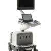


Philips Epiq 7 – Refurbished Ultrasound
EPIQ 7C is the new direction for premium ultrasound, featuring an exceptional level of clinical performance to meet the challenges of today’s most demanding cardiovascular practices.
Description
Philips Epiq 7 – Refurbished Ultrasound
More confidence in your diagnosis even for your most difficult cases
EPIQ 7C is the new direction for premium ultrasound, featuring an exceptional level of clinical performance to meet the challenges of today’s most demanding cardiovascular practices.
It’s our most powerful architecture ever applied to ultrasound imaging – touching all aspects of acoustic acquisition and processing, allowing you to truly experience ultrasound’s evolution to a more defi nitive modality.
Supported by our family of proprietary xMATRIX and PureWave transducers and our leading-edge Anatomical Intelligence, this platform off ers our highest level of premium performance.
Creating new realities, redefining clinical expectations
Philips nSIGHT Imaging is a totally new approach The Philips proprietary nSIGHT Imaging architecture introduces a totally new approach to forming ultrasound images. Unlike conventional systems that form the image line by line, nSIGHT creates images with superb resolution down to the pixel level.
Extraordinary architecture nSIGHT Imaging incorporates a custom multi-stage precision beamformer along with massive parallel processing. This proprietary architecture captures an enormous amount of acoustic data from each transmit operation and performs digital beam reconstruction along with mathematically optimized focal processing to create real-time images with exceptional resolution and uniformity.
The next generation of Color Flow Imaging
nSIGHT Imaging architecture incorporates new color Doppler technology that increases flow resolution, sensitivity, and frame rate. New proprietary flow algorithms produce exceptional vessel border delineation while preserving 2D imaging characteristics in color Doppler modes. New color map options allow enhanced visualization of flow for color-blind users.
New MicroCPA for exceptional small vessel visualization
In the past, obtaining flow information in small low-flow vascular structures has been a challenge. Now the EPIQ’s MicroCPA feature – visualization of low velocity micro circulation – is quick and simple, allowing for more diagnostic confidence when evaluating organ perfusion or small vascular beds.
Maximize extreme clinical capabilities
xMATRIX is our most leading-edge, versatile ultrasound transducer technology
No other premium ultrasound system can run the complete suite of the world’s most innovative ultrasound transducers. With the touch of a button xMATRIX offers all modes in a single transducer: 2D, 3D/4D, Live xPlane, Live MPR, MPR, Doppler, color Doppler, and CPA.
nSIGHT Imaging makes powerful xMATRIX technology even more so
Achieve ultra-thin 2D slices. Use Live xPlane imaging to create two full-resolution planes simultaneously, allowing you to capture twice as much clinical information in the same amount of time. Acquire near isovoxel resolution to reveal images from any plane within the volume. Now it’s all possible. Send 3D MPRs in the X, Y, and Z plane to any PACS system with MPR DICOM Export. Present superb, real-time 4D volume data in abdominal and obstetrical exams.
Greatly enhance the power of the X6-1 transducer for abdominal and OB applications
Now you can implement elevation compounding on the X6-1 with no frame-rate penalty for enhanced speckle reduction and contrast resolution at all depths. Use the X6-1 to perform real-time 4D imaging of the fetal heart or obtain a full 90° x 90° volume sweep of the liver in less than 0.25 seconds.
The power of PureWave to image technically difficult patients
PureWave crystal technology represents the biggest breakthrough in piezoelectric transducer material in 40 years. The pure, uniform crystals of PureWave are 85% more effi cient than conventional piezoelectric material, resulting in exceptional performance. This technology allows for improved penetration in diffi cult patients with a single transducer while maintaining excellent detail resolution and Doppler sensitivity.
Uniquely designed for elastography – revealing more definitive information on tissue stiffness
The EPIQ platform supports both strain and shear wave methods of elastography. Highly sensitive strain imaging can be used to rapidly assess relative tissue stiffness values across a variety of applications. Shear wave elastography utilizes a unique pulsing scheme to generate and detect the propagation speed of shear waves, providing an absolute measure of tissue stiffness. In addition, the EPIQ platform is designed to support the future of elastography including quantitative real-time shear wave imaging across a variety of transducers and applications.
Strain elastography
Philips strain elastography incorporates nanometer tissue strain tracking technology – a highly sensitive method of tracking tissue deformation requiring virtually no external compression for reproducible strain imaging results. Inherent patient physiologic movements provide the compression to generate the elastography image.
Shear wave elastography
ElastPQ uses ultrasound shear wave elastography to provide a non-invasive, reproducible and easily performed method of assessing tissue stiffness. A special pulse sequence technique using existing transducers produces shear waves in tissue and measures the propagation speed of the waves. Now tissue stiffness samples can be acquired during a routine ultrasound examination of the liver. According to the latest studies,1 using shear wave elastography may help reduce or avoid conventional liver biopsies. Many studies are suggesting that instead of a costly and painful biopsy procedure, an easy ultrasound exam becomes the routine method to assess liver disease status.
Revolutionize your contrast exam
Contrast-enhanced ultrasound (CEUS)* workflow is now seamlessly integrated into virtually any exam. EPIQ 7 provides immediate support of CEUS studies and exceptional performance across multiple agents and applications.
nSIGHT Imaging allows higher sensitivity to lower bubble concentrations while providing exceptional temporal resolution during critical wash-in/ wash-out phases. Among leading ultrasound manufacturers, Philips offers the world’s only live 3D images using contrast for general imaging with real-time 3D contrast data for dynamic clinical assessment.
Additional information
| Weight | 104 kg |
|---|---|
| Dimensions | 60 × 109 × 146 cm |
| Brand | |
| Condition | |
| Medical Specialty | Cardiology, Endocrinology, Gastroenterology, Neurosurgery, Obstetrics – Gynecology, Radiology, Rheumatology, Surgery, Urology, Veterinary |
| Availability | |
| Color | |
| Doppler | CW Color Doppler, PW Color Doppler, Real-time Doppler Auto Trace |
| Display Technology | |
| Image Quality | |
| Screen Dimensions | |
| Touch Panel | |
| Connectivity | |
| Print Type | Ethernet Color Printer, Thermal B/W Printer, USB Color Printer |
| Head ports | |
| System Portability |
Product Files
Product Files
Instruction Manual – Specifications
Declaration of Conformity
Compatible Probes
Compatible Probes
Convex
C8-5
C9-2
C5-1
Endocavity
C10-3v
C10-4ec
Linear
eL18-4
eL1-4 EM
L12-3
L15-7io
L12-5 50
L18-5
Phased Array
S5-1
S8-3
S12-4
Independent CW Doppler
D5cwc
D2cwc
D2tcd
TEE
S7-3t
S8-3t
xMatrix
X6-1
X5-1
X7-2
X7-2t TEE
X8-2t TEE
4D Probes
V6-2 4D Convex
3D9-3v
VL13-5
