
 Ελληνικά

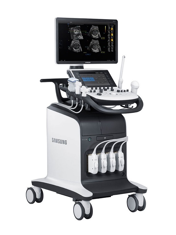
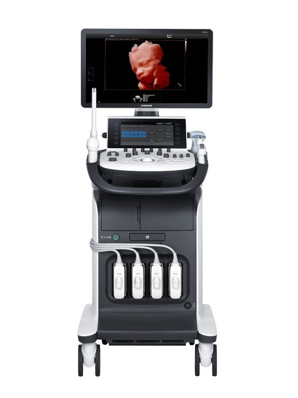
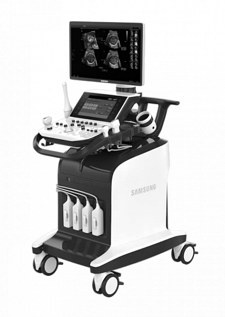
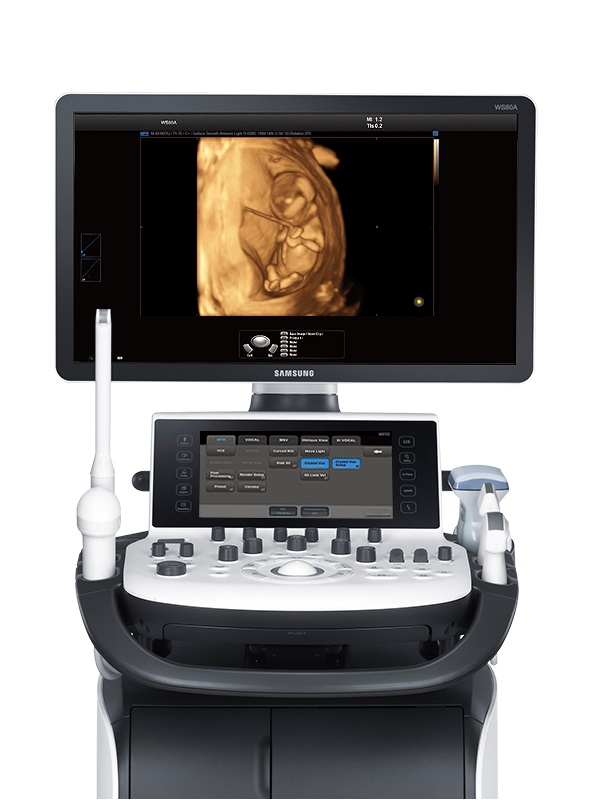
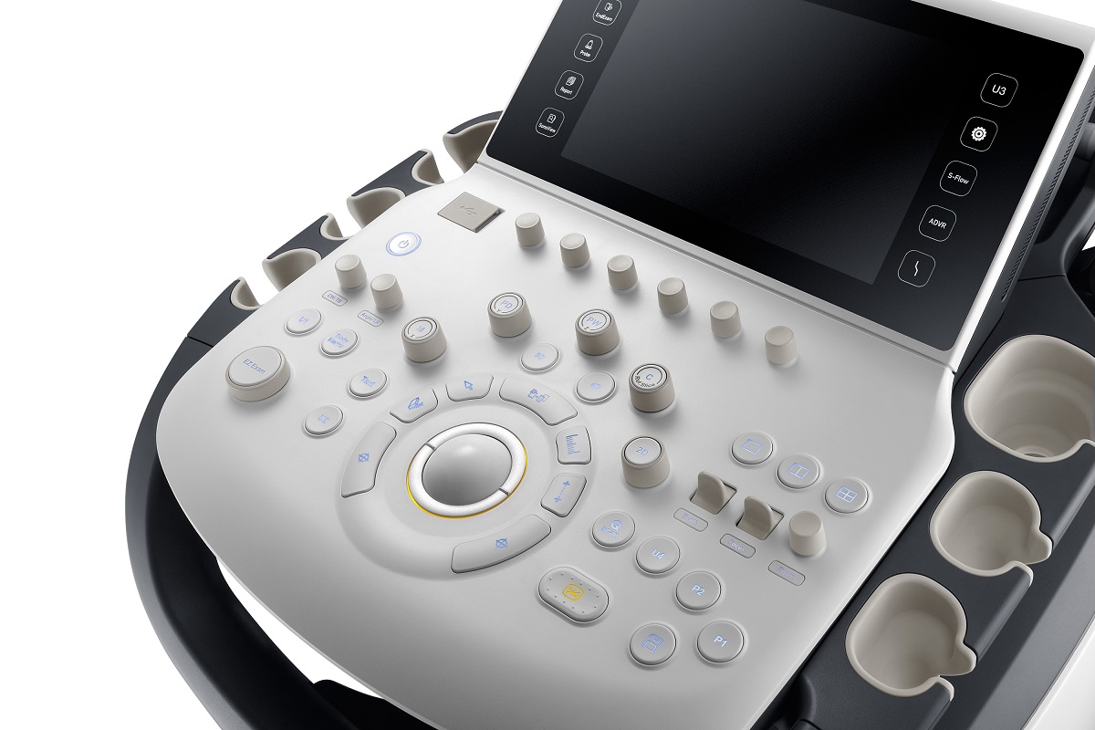
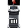
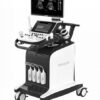
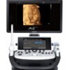
Samsung WS80A – Refurbished Ultrasound
The WS80A Elite is a high resolution premium system designed to meet the needs of women’s healthcare. WS80A Elite delivers exceptional image clarity by leveraging our S-Vision hybrid beamformer technology and S-Vue™ transducers. As pioneers in 3D/4D ultrasound, Samsung continues to advance volumetric imaging with Realistic Vue™ and 5D ultrasound.
Description
Samsung WS80A – Refurbished Ultrasound
The WS80A Elite is a high resolution premium system designed to meet the needs of women’s healthcare. WS80A Elite delivers exceptional image clarity by leveraging our S-Vision hybrid beamformer technology and S-Vue™ transducers. As pioneers in 3D/4D ultrasound, Samsung continues to advance volumetric imaging with Realistic Vue™ and 5D ultrasound.
Samsung Innovative Technologies
Hybrid Beamforming Technology
Samsung’s innovative hybrid beamformer technology is comprised of both advanced hardware and software, allowing for intricate digital programming, which better defines the shape of the ultrasound pulse. This provides more precise transmission and reception of the ultrasound signal, resulting in exceptional image clarity
S-Vue™ Transducer
In addition to the advanced beamforming capabilities, the WS80A Elite incorporates the next generation single crystal probe technology called S-Vue™ transducers. Employing a superior crystal design, S-Vue™ transducers provide more efficient piezoelectric properties, resulting in wider bandwidths for superior penetration and higher quality resolution on even the most challenging of patients.
ClearVision™
ClearVision™ is an advanced multi-filtering technology designed to decrease speckle, enhance border detection and display exceptional contrast resolution. ClearVision™ significantly enhances image clarity, providing more confident assessment of fetal anatomy.
S-Flow™
S-Flow™ is a highly sensitive Doppler technology utilizing both phase (directional) and amplitude data to ensure confident vascular documentation on even the smallest peripheral vessels.
MPI
MPI provides a quick and efficient method of performing fetal Myocardial Performance Index Doppler measurements. MPI also improves workflow during fetal heart examinations with an easy one-click operation.
ElastoScan™
ElastoScan™ technology facilitates an effective method for assessment and documentation of tissue stiffness. ElastoScan™ may prove an effective adjunct to conventional grayscale imaging, often providing more defined visualization of tumor margins.
5D ADVANCED APPLICATIONS
5D ultrasound is a suite of advanced applications designed to streamline workflow while enhancing reproducibility for a more consistent and confident clinical assessment. 5D is the logical advancement in volumetric ultrasound, addressing the needs of today’s women’s healthcare professionals. 5D semi-automated assessment of anatomy reduces exam time by locating standard image planes and anatomical structures including fetal heart, brain, long bones and nuchal translucency.
5D Heart (Fetal heart examination)
5D Heart is an innovative application designed to simultaneously display nine standard fetal cardiac views capturing one dynamic cardiac cycle. 5D Heart utilizes intelligent navigation technology to quickly assess fetal cardiac anatomy and simplify documentation of the fetal heart examination, thus reducing operator dependency, saving time and enhancing reproducibility.
5D CNS (Fetal brain measurement)
Using intelligent navigation, 5D CNS provides six measurements (BPD, HC, OFD, Cerebellum, Posterior Fossa, Atria of lateral ventricle) from three transverse views of the fetal brain to enhance measurement reproducibility and streamline workflow.
5D LB (Fetal long bone detection)
5D LB efficiently locates, displays and measures fetal long bones (Radius, Ulna, Humerus, Femur, Tibia and Fibula) from within the 3D dataset, streamlining workflow while enhancing measurement reproducibility.
5D NT (Nuchal translucency measurement)
5D NT intelligent navigation simplifies the NT measurement process by locating the precise mid-sagittal plane in a single step. Semi-automatic measurements are then performed to improve NT accuracy and reduce examination time.
5D Follicle (Follicle measurement)
5D Follicle identifies and measures multiple ovarian follicles for rapid assessment of follicular size and status during gynecology examinations.
SAMSUNG REALISTIC 3D IMAGING
Realistic Vue™
Realistic Vue™ displays high resolution 3D anatomy with exceptional detail and realistic depth perception. User determines the direction of a simulated light source, significantly enhancing clarity of surface detail and tissue parenchyma, as well as introducing intricate graduated shadows. Realistic Vue™ may also prove effective providing detailed 3D images for supportive documentation of pelvic anatomy. The ability to direct the simulated light source in combination with unique C-Plane images provides added value not attainable using conventional 2D imaging.
Additional information
| Weight | 105 kg |
|---|---|
| Dimensions | 79 × 55 × 140 cm |
| Brand | |
| Condition | |
| Medical Specialty | Cardiology, Endocrinology, Gastroenterology, Neurosurgery, Obstetrics – Gynecology, Radiology, Rheumatology, Surgery, Urology, Veterinary |
| Availability | |
| Color | |
| Doppler | CW Color Doppler, PW Color Doppler, Real-time Doppler Auto Trace |
| Display Technology | |
| Image Quality | |
| Screen Dimensions | |
| Touch Panel | |
| Connectivity | Analog Video Input/Output, DICOM, Ethernet, HDMI, USB 2.0, DVD External |
| Print Type | Ethernet Color Printer, Thermal B/W Printer, USB Color Printer |
| Head ports | |
| System Portability |
Product Files
Product Files
Instruction Manual – Specifications
Declaration of Conformity
Compatible Probes
Compatible Probes
Convex
CA1-7A
C2-6
SC1-6
CF4-9
Endocavity
VR5-9
E3-12A
Linear
LA3-16A
L5-13
L3-12A
Phashed Array
PE2-4
3D/4D
CV1-8A
V4-8
V5-9
LV3-14A
